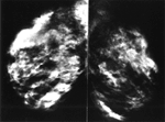
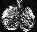
Digital Mammography
Conventional mammography uses X-rays to look for tumors or suspicious areas in the breasts. Digital mammography also uses X-rays, but the data is collected on computer instead of on a piece of film. This means that the image can be computer-enhanced, or areas can be magnified. Eventually, a computer could in certain appropriate situations, screen digital mammograms, theoretically detecting suspicious areas that human error might miss. There are both 2D and 3D forms of mammography available today.
In addition to "regular" mammograms, additional or spot views might be needed if a suspicious finding is noted on a screening mammogram (meaning no tumor was suspected by history nor by physical exam when the test was ordered).
US and MRI are other types of diagnostic tests that might be obtained to assess the breast.
Sometimes a biopsy is recommended in order to get a piece of tissue for the Pathologist to examine. This will determine if a tumor might be present.
In most every part of the body except for the breast, most biopsies find cancer or something else bad. In the breast, 80-90% of biopsies do not show any tumor. However, because imaging findings for cancer and non-cancerous abnormalities are similar, we have not yet found a way to improve on this low yield.
Breast cancer also occurs in men. So a man with a new chest mass should also see his doctor.