


CT Scans
A computed tomography scan (CT scan, also called a CAT scan) uses computer-controlled X-rays to create images of the body. However a radiograph and a CT scan show different types of information. Although an experienced radiologist can get a sense for the approximate three-dimensional location of a tumor from a radiograph, in general, a plain radiograph is two-dimensional.
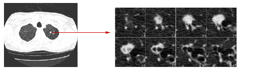
An arm or chest radiograph looks all the way through a body without being able to tell how deep anything is. A CT scan is three-dimensional. By imaging and looking at several three-dimensional slices of a body (like slices of bread) a doctor could not only tell if a tumor is present, but roughly how deep it is in the body. A CT scan can be three dimensional because the information about how much of the X-rays are passing through a body is collected not just on a flat piece of film, but on a computer.
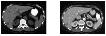
The data from a CT scan can be enhanced to be more vivid than a plain radiograph. For both plain radiographs and CT scans, the patient may be given a contrast agent to drink and/or by injection to more clearly show the boundaries between organs or between organs and tumors.
Recent technical advances in CT scanning dramatically increased its speed and effectiveness by a process called multi-slice or helical (spiral) scanning.
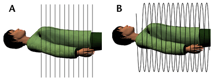
Conventional CT scans take pictures of slices of the body (like slices of bread). These slices are a few millimeters apart. The newer spiral (also called helical) CT scan takes continuous pictures of the body in a rapid spiral motion, so that there are no gaps in the pictures collected.
CT scanning is very common. Doctors will order a CT scan or MRI exam on most every patient with a suspect cancer.