


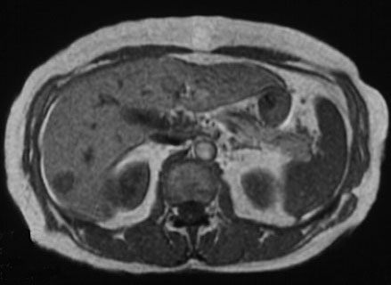
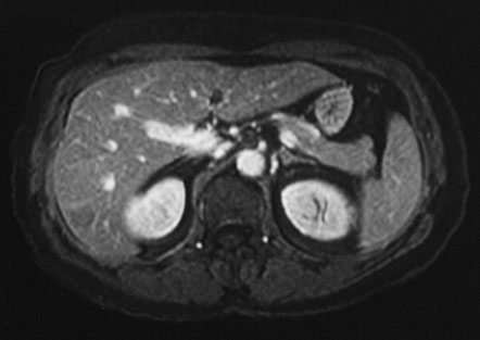
Magnetic Resonance Imaging (MRI)
Magnetic Resonance Imaging (MRI) uses radio waves in the presence of a strong magnetic field that surrounds the opening of the MRI machine where the patient lies to get tissues to emit radio waves of their own.
Different tissues (including tumors) emit a more or less intense signal based on their chemical makeup, so a picture of the body organs can be displayed on a computer screen. Much like CT scans, MRI can produce three-dimensional images of sections of the body, but MRI is sometimes more sensitive than CT scans for distinguishing soft tissues.
Many people know that MR scanners are noisy. Most every MR technologist has a big supply of ear plugs or an over the ear headphone for the patient to use. Some people get claustrophobic in the MR units. The Doctors and technologists are accustomed to helping patients deal with this.
MR is safe — so long as there is no metal in your body.
MR is safe for pregnant women and kids — there is no harmful radiation from MRI.